Which Structure Organizes The Mitotic Spindle During Cell Division . Centromere constricted region of a chromosome where sister chromatids are attached to one another and where the chromosome attaches to a spindle fiber. The mitotic spindle is a structure composed of microtubules which segregates chromosomes into the daughter cells during mitosis.
Review For Exam 2 Flashcards | Quizlet from quizlet.com
Which structure organizes the mitotic spindle during cell division? During mitosis, the spindle fibers are called the mitotic spindle.
Review For Exam 2 Flashcards | Quizlet
Intermediate filaments stable proteinaceous cytoskeletal elements that act as internal guy wires to resist mechanical (pulling) forces acting on cells Spindle fibers form a protein structure that divides the genetic material in a cell. What is a structure near the nucleus that functions during cell division?
Source: quizlet.com
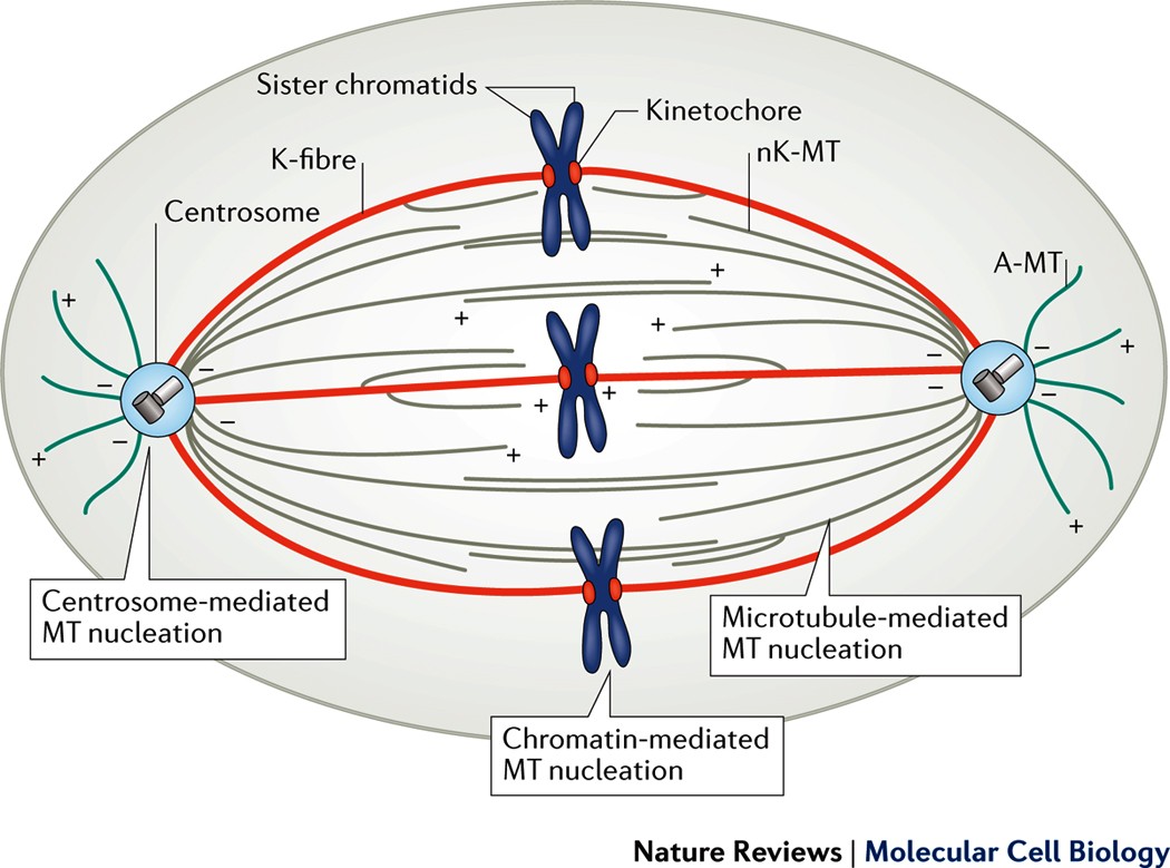
Overview of the mitotic spindle. They are formed from the centrosome, they are formed at the opposite poles during cell division and attach to chromosomes at the equatorial plate. They organize the cytoskeleton and direct formation of the spindle formed by the centrioles during cell division.
Source: www.nature.com
These spindle fibers pull the sister chromatids apart and ensure that one copy of each chromosome ends up in each daughter cell. Spindle fibers are the cellular structures that are used predominantly during cell division. The centrosome is an organelle that serves as a microtubule organizing center during cell division.
Source: quizlet.com
It is referred to as the mitotic spindle during mitosis, a process that produces genetically iden They are formed from the centrosome, they are formed at the opposite poles during cell division and attach to chromosomes at the equatorial plate. The mitotic spindle is a structure composed of microtubules which segregates chromosomes into the daughter cells during mitosis.
Source: www.researchgate.net
In eukaryotic cells, the mitotic apparatus is composed of two centrosomes and spindle microtubules ( figure 43.9. They organize the cytoskeleton and direct formation of the spindle formed by the centrioles during cell division. Spindle fibres are made up of tubulin proteins and rna.
Source: www.chegg.com
They are formed from the centrosome, they are formed at the opposite poles during cell division and attach to chromosomes at the equatorial plate. To coordinate cell division with chromosome segregation, the mitotic spindle controls cytokinetic events at the cell envelope. Mtocs have two main functions:
Source: en.wikipedia.org
Centrosomes always have assembled microtubules. The mitotic spindle is a structure composed of microtubules which segregates chromosomes into the daughter cells during mitosis. The centrosome is an organelle that serves as a microtubule organizing center during cell division.
Source: quizlet.com
During mitosis, the spindle fibers are called the mitotic spindle. Form the base of cilia and flagella. In eukaryotic cells, the mitotic apparatus is composed of two centrosomes and spindle microtubules ( figure 43.9.
Source: www.mdpi.com
In cell biology, the spindle apparatus (or mitotic spindle) refers to the cytoskeletal structure of eukaryotic cells that forms during cell division to separate sister chromatids between daughter cells. Centrioles are involved in the organizations of the mitotic spindle and in the completion of cytokinesis. The centrosomes, which migrate to opposite “poles” of the cell as the cell prepares for.
Source: quizlet.com
The organization of eukaryotic flagella and cilia and the organization of the mitotic and meiotic spindle apparatus, which separate the chromosomes during cell division. The mitotic spindle is a structure composed of microtubules which segregates chromosomes into the daughter cells during mitosis. During mitosis, the spindle fibers are called the mitotic spindle.
Source: www.nature.com
You can see that its dna has already. Spindle fibers are the cellular structures that are used predominantly during cell division. Centrosomes always have assembled microtubules.
Source: en.wikipedia.org
Centrosomes always have assembled microtubules. Intermediate filaments stable proteinaceous cytoskeletal elements that act as internal guy wires to resist mechanical (pulling) forces acting on cells It forms a protein structure that aids in the division of cells.
Source: www.frontiersin.org
The mtoc is a major site of microtubule nucleation and. Spindle fibers form a protein structure that divides the genetic material in a cell. During mitosis, the spindle fibers are called the mitotic spindle.
Source: www.facebook.com
The organization of eukaryotic flagella and cilia and the organization of the mitotic and meiotic spindle apparatus, which separate the chromosomes during cell division. Centrosomes always have assembled microtubules. A) 1 b) 2 c) 3 d) 5 e) 6 synthesis of carbohydrates and lipids occurs in the structure labeled a) 4.
Source: www.chegg.com
They are formed from the centrosome, they are formed at the opposite poles during cell division and attach to chromosomes at the equatorial plate. The mitotic spindle is a structure composed of microtubules which segregates chromosomes into the daughter cells during mitosis. Form the base of cilia and flagella.
Source: www.nature.com
Despite being central to the study of cell biology since its description in an animal cell by flemming in 1882 (), several aspects make the mitotic spindle one of the most challenging systems to grasp at quantitative and molecular levels.first, its size is immense, with the largest spindles measuring up to 60 μm in length ().second, it is complex, with.
Source: study.com
Spindle fibers form a protein structure that divides the genetic material in a cell. Cell organelle, existing in pairs, that occurs in the centrosome and may help organize a mitotic spindle for chromosome movement during animal cell division. Centrosomes always have assembled microtubules.
Source:
The spindle is necessary to equally divide the chromosomes in a parental cell into two daughter cells during both types of nuclear division: What is a structure near the nucleus that functions during cell division? Which structure organizes the mitotic spindle during cell division?
Source: www.mdpi.com
Form the base of cilia and flagella. To coordinate cell division with chromosome segregation, the mitotic spindle controls cytokinetic events at the cell envelope. It is referred to as the mitotic spindle during mitosis, a process that produces genetically iden
Source: www.chegg.com
In eukaryotic cells, the mitotic apparatus is composed of two centrosomes and spindle microtubules ( figure 43.9. Which structure organizes the mitotic spindle during cell division? You can see that its dna has already.
Source: www.jbc.org
Form the base of cilia and flagella. The mtoc is a major site of microtubule nucleation and. Direct the formation of the mitotic spindle during cell division;